
Wellness Edge Clinic
Punawales 2D echo & sonography center
for appointment call on : 9158442542


Services and history
Advanced Diagnostics: 2D Echo, Sonography, and Cardiac OPD in Punawale
When it comes to your health, precision in diagnosis ensures the right direction for treatment. At Punawale Multispeciality Hospital, our state-of-the-art diagnostic center brings advanced medical imaging and expert consultations under one roof. We provide high-quality, reliable, and swift diagnostic reporting to support families across Punawale and the entire Pimpri-Chinchwad (PCMC) region.
Our Core Specialized Services
1. Advanced 2D Echo Test Centre
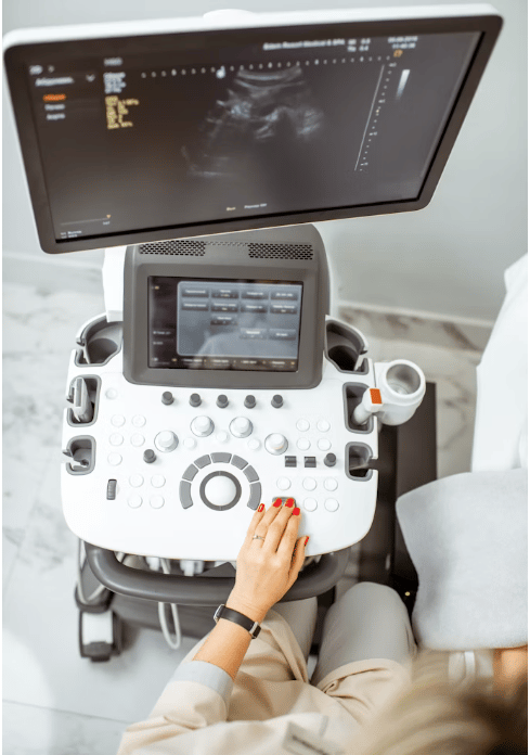
Evaluating your heart’s health requires clear, high-resolution imaging. Our 2D Echo (2D Echocardiogram) services provide a live, detailed look at your heart’s valves, chambers, and overall blood flow dynamics. This non-invasive ultrasound of the heart is critical for:
Detecting early signs of cardiovascular diseases.
Monitoring heart valve function and muscle strength.
Investigating unexplained chest pain, shortness of breath, or dizziness.
2. Comprehensive Cardiac OPD Services
Your heart deserves expert intervention. Our dedicated Cardiac OPD (Outpatient Department) is managed by senior cardiologists and physicians who offer personalized clinical evaluations. Whether you require regular hypertension monitoring, preventive heart check-ups, or expert guidance post-cardiac events, our OPD ensures you receive ethical and compassionate care without any transit delays.
3. High-Precision Sonography Centre
From routine abdomen checks to vital prenatal scans, our high-precision Sonography (Ultrasound) services utilize advanced imaging technology. Supervised by expert radiologists, we ensure safe, radiation-free, and highly accurate internal body imaging for:
Abdominal, pelvic, and KUB scans.
Obstetric and anomaly scans for pregnancy trac
People and culture
Our people are what make us unique. Rather than outsourcing our construction engineers from questionable outsourcing establishments, we provide them with an environment that supports professional growth.
We are strong believers in giving our employees a voice. Our teams are put together with the help of our resident psychologist to ensure maximum productivity and engagement.
Sonography & 2D Echo in Punawale:
Complete Guide to Ultrasound, Echocardiography, Doppler & Preparation
If you are searching for sonography in Punawale, ultrasound near me, 2D Echo in Punawale, echocardiography near me or diagnostic imaging in PCMC, understanding the different types of examinations can help you identify the investigation recommended by your doctor.
Sonography and 2D Echo both use ultrasound technology, but they have different purposes. General sonography can be used to examine various organs and soft tissues, while a 2D Echo is a specialized ultrasound examination of the heart.
Wellness Edge Clinic at Punawale Multispeciality Hospital provides sonography and 2D Echo services in Punawale.
What Is Sonography?
Sonography, commonly called ultrasound, is a diagnostic imaging technique that uses high-frequency sound waves to create images of structures inside the body.
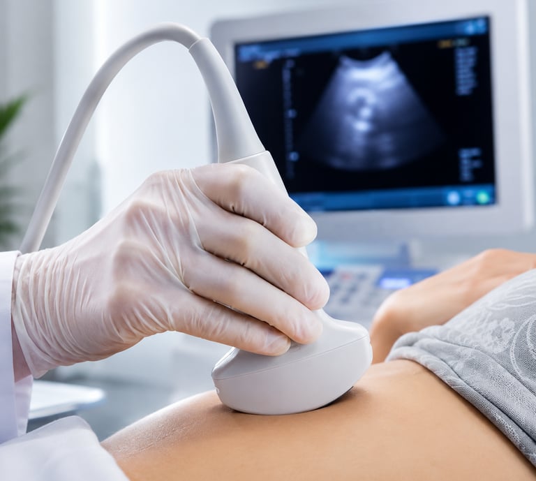
The examination uses a device called a transducer or probe. The probe sends sound waves into the body and receives returning echoes. The ultrasound system processes these signals to create images that can be viewed on a monitor.
Diagnostic ultrasound does not use ionizing radiation.
Depending on the examination prescribed by the doctor, ultrasound can be used to evaluate organs, soft tissues and blood vessels.
What Can Sonography Examine?
The specific area examined depends on the patient's symptoms and the doctor's prescription.
Common ultrasound examinations include:
Abdomen
Liver
Gallbladder
Pancreas
Spleen
Kidneys
Urinary bladder
Pelvis
Uterus and ovaries
Thyroid
Soft tissues
Blood vessels
Other areas requiring ultrasound evaluation
The appropriate examination depends on the clinical requirement.
Common Types of Sonography
Abdominal Sonography
An abdominal ultrasound can provide images of structures such as the liver, gallbladder, pancreas, spleen and kidneys.
Doctors may recommend abdominal sonography when investigating abdominal symptoms or assessing a suspected abnormality.
Depending on the examination, preparation instructions may include fasting.
Pelvic Sonography
Pelvic ultrasound provides imaging of structures in the pelvic region, including the uterus and ovaries.
It may be recommended when a doctor needs imaging information related to pelvic symptoms or gynecological concerns.
The exact type of pelvic ultrasound should be confirmed when booking.
Kidney and Urinary Tract Ultrasound
Ultrasound can be used to examine the kidneys and urinary bladder.
It may be recommended when evaluating certain urinary symptoms or suspected abnormalities.
Preparation requirements can vary depending on the examination.
Thyroid Sonography
Thyroid ultrasound provides detailed imaging of the thyroid gland.
Doctors may recommend it when evaluating thyroid enlargement, nodules or other structural changes.
Soft-Tissue Ultrasound
Ultrasound can also be used to evaluate selected superficial soft tissues.
The exact area and imaging technique depend on the clinical requirement.
Doppler Ultrasound
Doppler ultrasound uses ultrasound technology to provide information about blood flow.
It may be used when a doctor needs to evaluate circulation through particular blood vessels.
Depending on the body area being examined, Doppler may be performed as part of a broader ultrasound examination.
What Is a 2D Echo?
A 2D Echo, also called a two-dimensional echocardiogram, is a specialized ultrasound examination of the heart.
While general sonography can examine many parts of the body, echocardiography is specifically designed to provide moving images of cardiac structures.
A 2D Echo can provide information about:
Heart chambers
Heart valves
Heart muscle
Cardiac wall movement
Heart structure
Pumping function
Blood-flow patterns when Doppler techniques are used
The findings are interpreted in combination with the patient's symptoms, medical history and other investigations.
Why Is a 2D Echo Done?
A doctor may recommend an echocardiogram when information about the structure or function of the heart is clinically required.
Depending on the individual situation, an Echo may form part of the assessment of:
Breathlessness
Chest-related symptoms
Swelling
Abnormal heart sounds
Suspected valve abnormalities
Known cardiac conditions
Assessment of heart function
Follow-up of certain heart conditions
A 2D Echo should be performed when clinically appropriate and according to medical advice.
Disclaimer : The information provided in this article is intended for general educational and informational purposes only. It is not intended to replace professional medical advice, diagnosis, examination or treatment from a qualified healthcare professional.